
Ultrasound-Guided Sclerotherapy is a minimally invasive treatment for varicose and spider veins, using ultrasound to guide the injection of a sclerosant into problematic veins, promoting their closure.
Definition and Purpose
Ultrasound-Guided Sclerotherapy (UGS) is a minimally invasive medical procedure designed to treat varicose and spider veins. It involves using ultrasound imaging to guide the precise injection of a sclerosant solution into the affected veins. The primary purpose of UGS is to close off dysfunctional veins, restoring normal blood flow and alleviating symptoms such as pain, swelling, and cosmetic concerns. By leveraging ultrasound technology, the procedure ensures accurate delivery of the sclerosant, making it particularly effective for veins that are not easily visible or accessible. This treatment aims to improve both the functional and aesthetic outcomes for patients with venous insufficiency.
Historical Background and Development
Ultrasound-Guided Sclerotherapy (UGS) has evolved from traditional sclerotherapy, which dates back to the late 19th century. Initially, sclerotherapy involved injecting irritating solutions into varicose veins without imaging guidance, often resulting in variable success. The advent of ultrasound technology in the 20th century revolutionized the procedure by enabling precise visualization of veins. By the 1980s, ultrasound guidance became integral to sclerotherapy, particularly for deeper or less visible veins. This innovation improved accuracy, safety, and outcomes, making UGS a preferred treatment for varicose and spider veins. Today, UGS is recognized as a highly effective, minimally invasive approach for venous insufficiency.

The Procedure of Ultrasound-Guided Sclerotherapy
Ultrasound-Guided Sclerotherapy involves using ultrasound imaging to precisely guide the injection of a sclerosant into targeted veins, ensuring accurate delivery and effective treatment of varicose or spider veins.
Preparation for the Procedure
Preparation for Ultrasound-Guided Sclerotherapy involves avoiding blood thinners, wearing loose clothing, and fasting if required. Patients should remove lotions or oils from the skin and avoid tight garments. The procedure typically takes 30-45 minutes, during which the patient lies on their back or in a position that enhances vein visibility. The healthcare provider may also review medical history and allergies to ensure safety. Proper preparation ensures the procedure is performed efficiently and effectively, minimizing risks and optimizing outcomes.
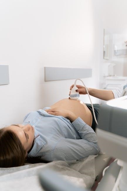
Role of Ultrasound in Guiding the Treatment
Ultrasound technology plays a crucial role in guiding the sclerotherapy procedure by providing real-time imaging of the veins. This allows the physician to precisely locate and visualize the target veins, especially those hidden beneath the skin. The ultrasound ensures accurate placement of the sclerosant, minimizing the risk of complications. By enabling visualization of vein anatomy and blood flow, ultrasound enhances the effectiveness of the treatment and improves patient outcomes. This advanced imaging guidance is particularly beneficial for veins that are difficult to access or visualize, making the procedure safer and more precise compared to traditional sclerotherapy methods.
Administration of the Sclerosant
During ultrasound-guided sclerotherapy, the sclerosant is administered directly into the targeted veins under real-time ultrasound visualization. A small needle is used to inject the sclerosant, which can be in liquid or foam form, depending on the vein size and location. The ultrasound ensures precise placement, reducing the risk of injecting surrounding tissues. The sclerosant works by irritating the vein lining, causing it to swell, close off, and eventually disappear. The procedure is typically performed with the patient lying down, and the process is repeated for multiple veins if necessary. This targeted approach minimizes discomfort and enhances treatment efficacy.
Advantages of Ultrasound-Guided Sclerotherapy
Ultrasound-guided sclerotherapy is highly effective, minimally invasive, and offers precise targeting of veins. It reduces recovery time, minimizes complications, and provides superior cosmetic results compared to traditional methods.
Effectiveness in Treating Varicose Veins
Ultrasound-guided sclerotherapy is highly effective in treating varicose veins, offering significant improvement in both symptoms and appearance. By using ultrasound imaging, the procedure ensures precise delivery of the sclerosant directly into the affected veins, leading to their closure and eventual absorption by the body. Clinical studies demonstrate high success rates, with many patients achieving long-term relief from venous insufficiency. The use of foam sclerotherapy, guided by ultrasound, is particularly effective for larger varicose veins, providing a minimally invasive alternative to surgery. Patients often report improved quality of life and satisfaction with the aesthetic and functional outcomes of the treatment.
Minimally Invasive Nature
Ultrasound-guided sclerotherapy is a minimally invasive procedure that eliminates the need for major surgery. It involves inserting a small needle into the vein under ultrasound guidance, reducing trauma to surrounding tissue. This approach minimizes discomfort, scarring, and recovery time compared to traditional surgical methods. Patients typically experience mild discomfort during the procedure, and no general anesthesia is required. The minimally invasive nature also lowers the risk of complications, such as infection or prolonged downtime, making it a safer option for many individuals. This technique is particularly advantageous for treating veins that are difficult to access or visible only with ultrasound imaging.
Improved Accuracy with Ultrasound Guidance
Ultrasound guidance significantly enhances the accuracy of sclerotherapy by providing real-time visualization of the vein structure. This technology allows practitioners to precisely locate and target veins that are deep or not visible to the naked eye. By using ultrasound, the sclerosant is delivered directly into the affected vein, minimizing the risk of misplacement and reducing potential complications. The enhanced visualization ensures that the treatment is more effective and safer, especially for veins that are challenging to access. This level of precision is a major advancement over traditional sclerotherapy, making the procedure more reliable and efficient for patients with varicose or spider veins.
Indications for Ultrasound-Guided Sclerotherapy
Spider Veins
Spider veins are small, visible veins near the skin’s surface, often appearing as red or purple clusters. Ultrasound-guided sclerotherapy effectively treats these unsightly veins when they are too small for other methods.
Varicose Veins
Varicose veins are enlarged, twisted veins that often appear swollen and bulging beneath the skin, typically in the legs. They result from improper blood flow and vein valve dysfunction. Ultrasound-guided sclerotherapy is highly effective for treating varicose veins, especially those that are not easily visible or accessible. The procedure involves using ultrasound to precisely locate the problematic veins and guide the injection of a sclerosant solution, which causes the vein to close and eventually disappear. This minimally invasive approach reduces the risk of complications compared to traditional surgery and offers significant improvement in symptoms such as pain, swelling, and cosmetic concerns. It is particularly beneficial for deeper veins that are difficult to treat with surface-level methods, providing both therapeutic and aesthetic benefits with a high success rate and minimal downtime.
Spider veins are small, dilated blood vessels that appear as red or purple lines on the skin’s surface, often resembling a spider’s web. They typically occur on the legs and face due to increased pressure or weakened vein walls. Ultrasound-guided sclerotherapy is an effective treatment for spider veins, especially when they are too small or deeply located for traditional sclerotherapy. The procedure uses ultrasound to visualize the veins, allowing precise injection of a sclerosant solution that closes the vein. Over time, the body absorbs the treated vein, reducing its visibility. This method is minimally invasive, offering both aesthetic improvement and relief from associated discomfort, with minimal recovery time compared to surgical options.
Chronic Venous Insufficiency
Chronic Venous Insufficiency (CVI) occurs when veins struggle to return blood to the heart due to faulty valves, leading to pooling and increased pressure. This condition often results in varicose veins, swelling, and discomfort. Ultrasound-guided sclerotherapy is an effective treatment for CVI, particularly for deeper veins that are difficult to address with surface treatments. The procedure involves using ultrasound to guide the injection of a sclerosant into the affected veins, causing them to close and redirect blood flow to healthier veins. This minimally invasive approach reduces symptoms like pain and swelling, improving overall venous function and quality of life for patients with CVI.
Contraindications and Risks
Contraindications include pregnancy, active cancer, or deep vein thrombosis. Risks involve allergic reactions, skin discoloration, or nerve damage, requiring careful patient screening and skilled administration.
Contraindications for the Procedure
Ultrasound-guided sclerotherapy is not suitable for everyone. Contraindications include pregnancy, breastfeeding, active cancer, or deep vein thrombosis. Patients with allergies to sclerosants or severe systemic diseases should avoid this treatment. Additionally, those with acute infections, severe peripheral artery disease, or pacemakers may not be candidates. A thorough medical history and examination are essential to ensure safety and eligibility for the procedure. Consultation with a healthcare provider is crucial to determine if ultrasound-guided sclerotherapy is appropriate for individual cases.
Possible Side Effects and Risks
Ultrasound-guided sclerotherapy is generally safe, but potential side effects include mild pain, swelling, or bruising at the injection site. Temporary skin discoloration or itching may occur. Rarely, more serious complications like blood clots, allergic reactions, or nerve damage can happen. Infection or scarring are uncommon but possible. Patients should discuss their medical history with their provider to assess individual risks. While the procedure is well-tolerated by most, it is not entirely without complications. Close follow-up and adherence to post-procedure care can minimize these risks and ensure optimal outcomes.
Post-Procedure Care and Recovery
After ultrasound-guided sclerotherapy, patients should avoid prolonged standing, wear compression stockings, and elevate legs to reduce swelling. Avoid heavy exercise, hot baths, and tight clothing temporarily.
Immediate Aftercare
Following ultrasound-guided sclerotherapy, patients are advised to avoid strenuous activities for 24-48 hours. Compression stockings should be worn as directed to reduce swelling and promote healing. Mild discomfort or bruising at injection sites is common but temporary. Ice packs can be applied to alleviate swelling. Patients should avoid hot baths, saunas, or direct sunlight for 2-3 days. Light walking is encouraged to prevent blood clots. Avoid heavy lifting or bending. Any unusual symptoms, such as severe pain or swelling, should be reported to the healthcare provider promptly. Patients can typically resume normal activities within a few days, with full recovery expected within weeks.
Long-Term Recovery and Outcomes
Most patients experience significant improvement in varicose veins within 3-6 months post-procedure. The treated veins typically close and disappear, reducing symptoms like pain and swelling. Long-term outcomes show high success rates, with over 80% of patients achieving lasting results. Some may require retreatment for residual veins. The procedure improves both aesthetic appearance and functional symptoms, enhancing quality of life. While ultrasound-guided sclerotherapy is effective, it does not prevent new varicose veins from forming. Ongoing vein health practices, such as regular exercise and compression stockings, may be recommended to maintain results and prevent recurrence. Overall, it offers a durable solution for many patients with varicose veins.
Ultrasound-Guided Sclerotherapy is a highly effective, minimally invasive treatment for varicose and spider veins. By combining sclerosant injection with real-time ultrasound imaging, it offers precise targeting of problematic veins, leading to significant symptom relief and aesthetic improvement. The procedure is well-tolerated, with minimal downtime, making it an attractive alternative to surgery. Long-term outcomes demonstrate lasting results for many patients, though retreatment may occasionally be needed. Overall, it represents a major advancement in vein treatment, providing both functional and cosmetic benefits with a strong safety profile;